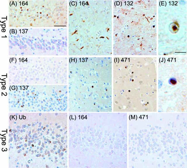
FIGURE 2.
Immunoreactivity profile for newly generated antibodies in FTLD-U cases with different patterns of inclusion pathology. A–E: Type 1: mAbs 164 and 132 label numerous cytoplasmic and neuritic inclusions in dentate gyrus (A; case 4, mAb 164), frontal cortex (C; case 4, mAb 164), and striatum (D; case 11, mAb 132) as well as intranuclear inclusions in striatal neurons (E; case 11, mAb 132). Inclusions in the dentate gyrus of a type 1 case are not labeled with mAb 137 (B; case 4, mAb 137). F–J: Type 2: Cytoplasmic inclusions are labeled with mAbs 137 and 471 in dentate gyrus (G; case 13, mAb 137), frontal cortex (H; case 13, mAb 137), and striatum (I; case 14, mAb 471). Intranuclear neuronal inclusion in temporal cortex labeled with mAb 471 (J, case 14). Inclusions in the dentate gyrus of a case with type 2 pathology are negative with mAb 164 (F, case 13). K–M: Type 3: Ubiquitin-positive inclusions in the dentate gyrus (K) of case 35, type 3, are negative for mAbs 164 (L) and 471 (M). Scale bars: 50 μm (A–D, F–I, K–M); 10 μm (E, J).

