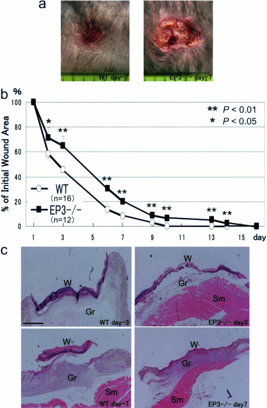
FIGURE 1.
Delayed wound healing in EP3 receptor knockout mice. Surgical wounds were made on the backs of EP3 receptor knockout mice (EP3−/−) and their WT counterparts, and wound closure was determined as described in Materials and Methods. a: Typical appearance of wounds in EP3−/− and WT at day 7. The original diameter of the wounds was 8 mm. One division on the scale below the wound represents 1 mm. b: Time course of wound closure in EP3−/− and WT. Data are means ± SEM for the indicated number of mice. *P < 0.05, and **P < 0.01 versus WT mice (analysis of variance). c: H&E staining was used for wound tissues including granulation tissues from EP3−/− and WT. Tissues were fixed at days 3 and 7. W, wound; Gr, granulation tissue; Sm, skeletal muscle. Scale bar = 1 mm.

