FIGURE 3.
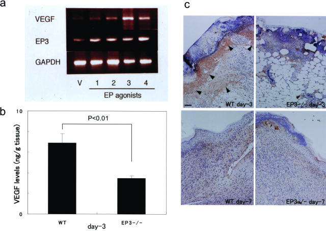
Expression of VEGF in sponge granulation tissues in WT mice stimulated with selective EP agonists and in wound granulation tissues in EP3 receptor knockout mice. a: VEGF expressions in sponge granulation tissues in WT mice stimulated with selective EP agonists for 2 weeks. EP receptor subtype-selective agonists, ONO-DI-004, ONO-AEI-257, ONO-AE-248, and ONO-AEI-329, which were specific to EP1, EP2, EP3, and EP4, respectively,18 were topically injected (15 nmol/sponge, twice a day) into the sponges implanted in the subcutaneous tissues of WT mice. RT-PCR was performed as described in the text. b: Immunoreactive VEGF levels, determined by ELISA, in the wound granulation tissues in EP3−/− mice and WT mice at day 3. VEGF protein was not detected in normal skin tissue in WT mice (data not shown). VEGF expression was detected in wound tissue in WT mice (left) but was reduced in EP3−/− mice (right). To test the significance of difference, t-test was used. c: Typical results of VEGF immunostaining in the wound granulation tissues in EP3−/− and WT at days 3 and 7. VEGF-positive cells were markedly accumulated in the wound granulation tissues in WT mice at day 3 (brown stains indicated by arrowheads). Scale bar = 100 μm.

