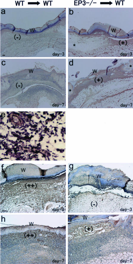
FIGURE 5.
Typical results of immunostainings of β-galactosidase (a–d) and VEGF (f–i) and of EP3 in situ hybridization (e) in the wound granulation tissues in WT mice transplanted with BM cells either from EP3−/− mice (EP3−/−→WT) or from WT mice (WT→WT) at day 3 (a, b, e–g) and at day 7 (c, d, h, and i). β-Galactosidase-positive cells accumulated in the wound granulation tissues in WT mice transplanted with BM cells from EP3−/− mice (b and d, ++). WT mice receiving WT BM cells did not show β-galactosidase-positive stains in the wound tissues (a and c). The uninjured lesions around the wounds introduced in WT mice transplanted with BM cells from EP3−/− mice did not exhibit β-galactosidase (b and d, asterisks). EP3 mRNA was detected in granulation tissues just beneath the wound in WT mice receiving WT BM cells (e, high magnitude). VEGF immunoreactivity was mainly seen in granulation tissues in WT mice receiving WT BM cells (f and h), whereas no marked accumulation of VEGF-positive cells was seen in WT mice implanted with EP3−/− cells (g and i). W indicates surgical wound in recovery stage. Scale bars = 100 μm (a, f); 25 μm (e).

