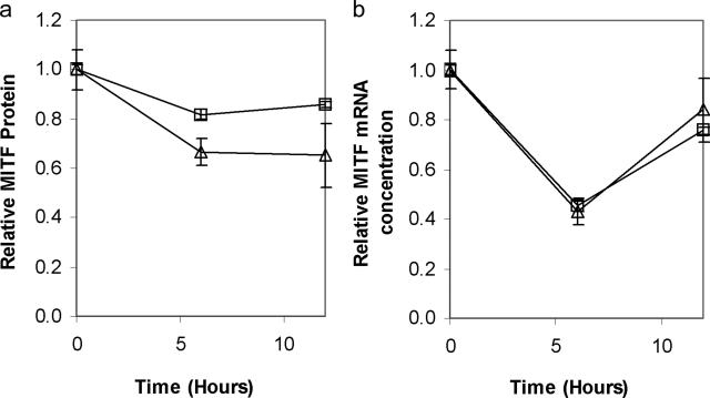
Figure 3.
4-TBP exposure results in reduced expression of both MITF RNA and protein in normal melanocytes. Cells from two separate normal human melanocyte lines were treated with 200 μmol/L 4-TBP. Samples were harvested over a 12-hour period. a: Cell lysates were prepared and normalized for protein content, then subjected to Western blot analysis followed by densitometry using an antibody against MITF. Total (both un- and phosphorylated) MITF protein content decreased within a 6-hour period. b: Cells from each of the two lines were also harvested for RNA extraction. Real-time PCR was performed in triplicate. MITF RNA concentrations were normalized to glyceraldehyde-3-phosphate dehydrogenase expression and are shown relative to MITF at 0 hours. Error bars show SD of the triplicates. Levels of MITF RNA were reduced to below 50% 6 hours after treatment with 4-TBP.

