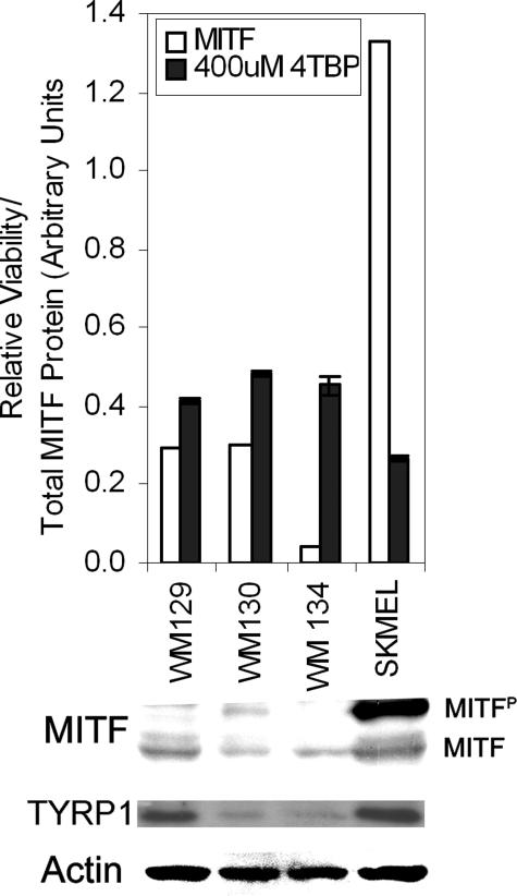
Figure 5.
Melanoma sensitivity to 4-TBP correlates with expression of MITF. Melanoma cells were either harvested for protein extraction or plated to test sensitivity to 4-TBP. Cells were attached for 48 hours and then dosed with 400 μmol/L 4-TBP for 72 hours. The proportion of viable cells was determined and is shown relative to cells treated with vehicle alone. Densitometry was performed following Western blot analysis for MITF, Tyrp1 (Pep1), and actin, total MITF protein is expressed as the sum of band densities for un- and phosphorylated MITF normalized to actin. SK-MEL188 that expressed the most MITF, and in the potent phosphorylated form, as well as Tyrp1, was significantly more sensitive than each of the other lines, which express less MITF (P < 0.05). Error bars indicate SD of triplicates within a representative experiment.

