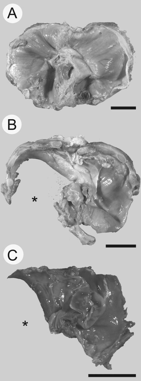
Figure 2.
Photomicrographs of human diaphragms. A normal diaphragm at 35 weeks of gestation (A) is shown for comparison against a diaphragm isolated from a fetus at 34 weeks of gestation (B) with a large left-sided diaphragm defect (*) and a diaphragm isolated from a 30-week fetus (C) also with a left-sided defect (*). Diaphragms are oriented such that the top of the image is anterior and the bottom of the image is posterior. Scale bar = 2 cm.

