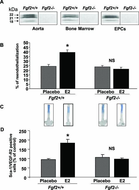
Figure 1.
Effect of estradiol (E2) on the reendothelialization process (3 days after injury) on EPC mobilization in ovariectomized Fgf2+/+ and Fgf2−/− mice treated (E2) or not (placebo) with E2. A: FGF-2 protein expression in aorta, bone marrow, and EPCs. B: The percentage of reendothelialization represents the ratio of the area remaining deendothelialized at day 3 on the area initially deendothelialized (eight mice in each group). *P < 0.05 versus respective control. C: Representative Evans blue dye staining of carotid 3 days after injury. D: EPC levels in peripheral blood were assessed from ovariectomized FGF2+/+ and FGF-2−/− mice treated (E2) or not (placebo) with E2 by FACS analysis to quantify Sca-1/VEGF-R2-positive cells (eight mice in each group, *P < 0.05 versus respective placebo).

