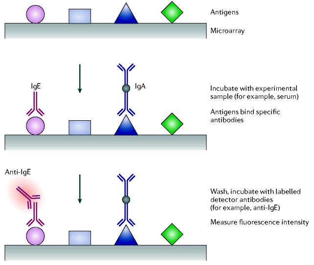
Figure 2.
Schematic representation of antigen or peptide capture arrays. Antigens are printed on the array, which is then incubated with an experimental sample containing antibodies. Binding of the antibodies to the antigens can then be detected by binding a secondary, anti-immunoglobulin (a read-out antibody) that has been fluorescently labelled (red halo). Ig, immunoglobulin.

