
Figure 8.
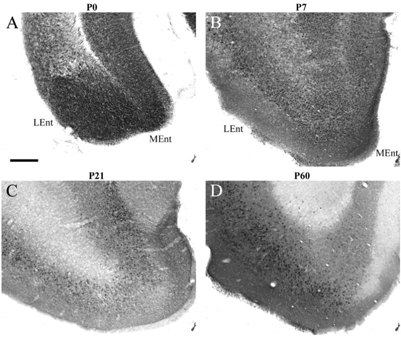
RGS4-GFP expression in the P0-P60 entorhinal cortex. Expression of RGS4-GFP in the entorhinal cortex at P0 (A), P7 (B), P21 (C), P60 (D) following immunohistochemical visualization with an anti-GFP antibody and DAB staining. Note the decreased expression of RGS4 in the medial entorhinal cortex at P60. LEnt, lateral entorhinal cortex; MEnt, medial entorhinal cortex. Scale bar in A = 200 μm for all images.