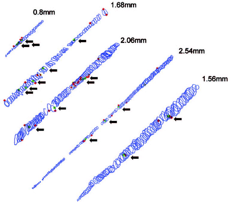
Figure 5.

Reconstructions of five myofibers from serially sectioned (12 μm) resected muscle immunostained for BrdU and dystrophin. Each circle represents a single cross-sectional area. Red: BrdU-positive satellite cells; green: BrdU-positive myonuclei. Arrows: each BrdU-positive myonucleus. The total length is indicated for each reconstructed myofiber.
