
FIGURE 6.
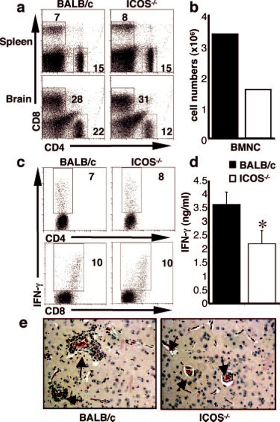
ICOS-/- mice have less inflammation at the chronic stage of T. gondii infection. BALB/c and ICOS-/- mice were infected with T. gondii, and after 4 wk of infection, BMNC numbers were counted (b), and these and splenic cells were incubated with Abs to CD4 and CD8 (a). BMNC were cultured for 48 h with parasite Ag (sTAg) and stained for intracellular IFN-γ (c) as described in Materials and Methods. Live and cell-specific gates were applied. Supernatants were harvested and measured for IFN-γ (d) by ELISA. Paraffin sections of brains from naive and infected BALB/c and ICOS-/- mice were stained with H+E (e); arrows point to blood vessels to highlight the difference in inflammation, in particular in the perivascular infiltrate. Data shown are averages of at least three mice and are representative of at least three individual experiments. Significance where p < 0.05 is illustrated by *.