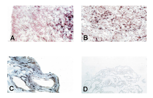
Figure 2.

In situ hybridization on rheumatoid arthritis synovial tissue shows only negligible expression of PTEN in the lining layer (A) but abundant expression in the sublining layer (A, B). Normal synovium consisting of only two to three cell layers of synovial cells showed clear staining, both in the most superficial layers and in deeper regions (C). The sense probe gave no specific staining (D).
