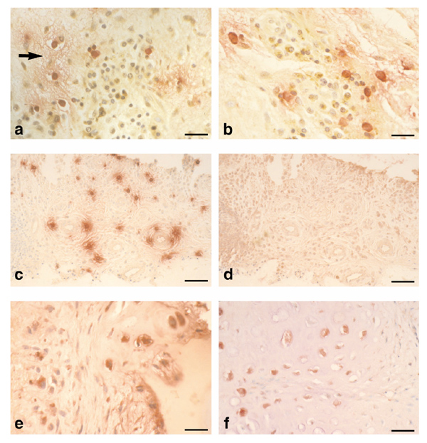
Figure 1.

Immunolocalisation of mast cell tryptase, tumour necrosis factor (TNF)-α, interleukin (IL)-1α and IL-15 in the rheumatoid lesion. (a) Micrograph showing mast cell tryptase (red) with extracellular staining indicative of mast cell (MC) activation associated with localized oedema (arrow) and TNF-α expression (brown) by a proportion of mononuclear cells. (b) Micrograph showing mast cell tryptase (red), local MC activation and associated mononuclear cells stained for IL-1α (brown). (c) Low power micrograph showing distribution of mast cells (tryptase, red) and their degranulation in rheumatoid synovial tissue. (d) Consecutive section to (c) stained for IL-15. Note the absence of IL-15 from MC activation sites. (e) Micrograph of cartilage-pannus junction showing both extracellular and intracellular staining for IL-15 (red). Note the microfocal nature of cytokine production by a minority of cells. (f) Micrograph of rheumatoid lesion showing chondrocytic cells stained for IL-15 (red) with little evidence of cytokine production by the overlying pannus tissue (to right of micrograph). Bars: (a) and (b) 35 μm; (c) and (d) 120 μm; (e) 35 μm; and (f) 45 μm.
