
Figure 3.
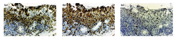
Immunohistological analysis of RA synovial membrane. Serial tissue sections were stained with (a) antibody to A-SAA, (b) anti-CD68 and (c) IgG. A-SAA-positive cells are seen in the surface of the lining layer and in the perivascular areas. Some vascular endothelial cells are weakly positive (a). CD68-positive cells are seen throughout the lining layer and in the perivascular areas of the sublining layer. Vascular endothelial cells are negative (b). No staining was observed in the control sections (c).