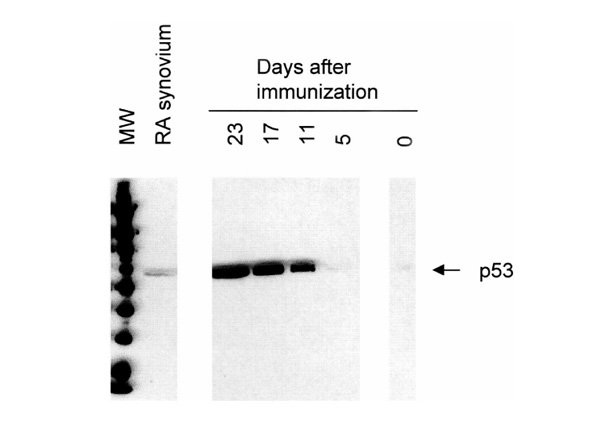
Figure 2.

Western blot analysis showing immunoreactive p53 in pooled joint extracts of rats with AA and pooled synovial tissue samples of patients with RA. Expression of p53 gradually increased from days 0-23 in rat AA (see also Table 1). Overexpression on day 23 (173 arbitrary units) was markedly higher than p53 levels in RA synovium (32 arbitrary units).
