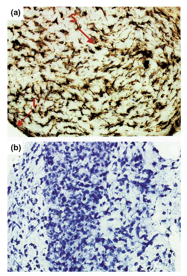
Figure 3.

Representative synovial tissue from a rat with adjuvant arthritis on day 23, showing marked p53 overexpression [(a), indicated by arrows]. Both cytoplasmic and nuclear staining was noted in the intimal lining layer (L) and in the synovial sublining (S). Staining was absent in the negative control section (b). Monostaining peroxidase technique with tyramine enhancement counterstained with Mayer's hemalum. Original magnification 400x.
