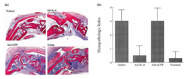
Figure 2.

Histological analysis of the effect of local Ad-mIL-4 treatment in CIA. (a) Ankle joints of mice were isolated from CIA and same-aged normal DBA mice 28 days after adenovirus injection. Ankle joint tissues were stained with hematoxylin and eosin and showed 100× magnification. Particles of Ad-IL-4 or Ad-eGFP (5×10⁸) were injected into ankle joints of mice with established CIA. (b) The joint tissue sections were evaluated in a blinded manner and scored as follows: 1, synovial cell proliferation, synovial hypertrophy with villus formation and/or fibrin deposition; 2, inflammation, synovitis and/or generalized inflammation; 3, cartilage disruption, chondrocyte degeneration and/or ruffling of cartilage surface and/or dystrophic cartilage; and 4, joint destruction, cartilage erosion with abundant inflammation and pannus formation with bone erosion. A total of five joints per group were evaluated by at least two individuals in a blinded manner.
