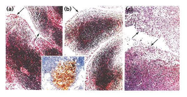
Figure 3.

Histopathology (haematoxylin and eosin) and immunohistochemistry (double staining: indirect immunoperoxidase and alcaline phosphatase in insert of parts a and b) of rheumatoid synovial tissue from three different anatomical locations of the RA patient. These are as follows: (a) right peroneal tendon sheath; (b) left peroneal tendon sheath with inserted figure showing Ki-M4 positive FDC network (brown) surrounded by CD20+ B lymphocytes (blue), representing a germinal center; and (c) synovial membrane from the right cubita. Arrows indicate enlarged synovial intima (original magnification 350×).
