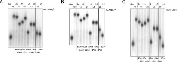
FIGURE 2.
Electrophoretic analysis of K-turn folding. Radioactively 5′-32P-labeled 65-bp DNA/RNA/DNA duplex species with a central Kt-7 sequence were electrophoresed in 15% polyacrylamide gels in 90 mM Tris.borate (pH 8.3) plus 500 μM (A) or 2 mM (B) Mg2+, or 10 μM Co(III) hexammine (C) salts. Phosphorimages of the dried gels are shown. The simple duplex species was loaded at the left, followed by the unmodified Kt-7 sequence. These two species define the range of electrophoretic mobilities. Single 2′-deoxyribose variants were electrophoresed, indicated by the position of the modification in the K-turn nomenclature (written above tracks) or in the Kt-7 numbering system (below tracks). The original gels included three tracks not related to the experiments discussed here between the tracks containing L3 and 2b variants; these have been removed for clarity and the position indicated by the bold line. The gray lines have been added to aid identification of different tracks. Two versions of the L1 modification were used, with the natural guanine (dG94) and variant adenine (dA94).

