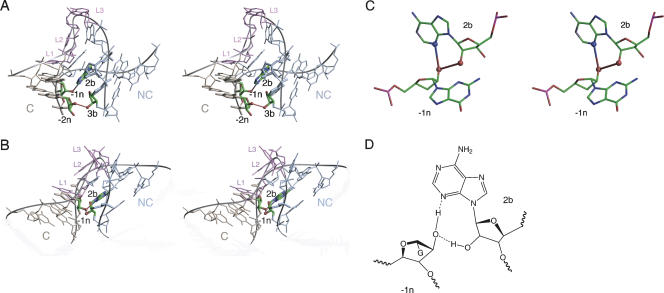
FIGURE 4.
Interactions between the C and NC helices found in crystal structures of Kt-7 (Klein et al. 2001), box C/D (Moore et al. 2004), and U4 (Vidovic et al. 2000) K-turns. (A,B) Parallel-eye stereo view of Kt-7 (A) and box C/D (B) K-turns looking down into the minor groove interface between the C and NC helices, with the bulge shown at the top. The C helix is colored gold, NC colored blue, and bulge nucleotides are colored purple; this scheme has been used in most graphics images in this article. The nucleotides participating in hydrogen bonding interactions are highlighted, and the bond drawn in. In Kt-7 (A) hydrogen bonds are seen between the 2′-OH groups of nucleotides −2n and 3b, and between the 2′-OH of −1n and N1 of adenine 2b. In box C/D (B) there is no interaction between nucleotides −2n and 3b. However, there are hydrogen bonds between the 2′-OH of −1n and N3 and 2′-O of adenine 2b. (C) Close-up view of the hydrogen bonding between the 2′-OH of −1n and N3 and 2′-O of adenine 2b in the U4 K-turn. (D) The chemical structure of the hydrogen bonding pattern between the 2′-OH of −1n and N3 and 2′-O of adenine 2b in box C/D and U4 K-turns.

