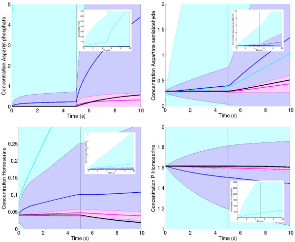
Figure 6.
Simulation results for threonine model. The refined parameter distributions lead to better predictions of the dynamic behaviour. Top left: simulated time series for aspartyl-phosphate. The curve from the true model is shown by black squares. After five minutes, the substrate aspartate is shifted to a higher concentration, leading to an increase of aspartyl-phosphate. Each parameter ensemble creates a distribution of simulation results: areas represent the standard deviations, the colours represent prior (light blue), kinetics-based posterior (dark blue) and metabolics-based posterior (purple). Inset: other scaling to show the relative spread of prior and first posterior. Other diagrams: time series for the remaining metabolites aspartate semialdehyde (top right), homoserine (bottom left), and p-homoserine (bottom right).

