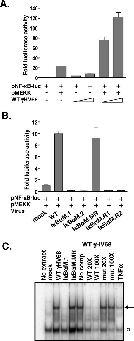
Figure 2. γHV68-IκBαM Inhibits NF-κB Signaling in Fibroblasts.
(A) NIH 3T12 fibroblasts were infected with WT virus at an MOI of 1 or 10 for 6 h and then transfected with a NF-κB–responsive luciferase reporter construct and, where indicated, a plasmid expressing MEKK1. Data are shown as the mean fold activation of the NF-κB promoter over mock infected samples ±SD of triplicate wells.
(B) NIH 3T12 cells were infected with the indicated viruses at an MOI of 10 for 6 h and then transfected with the NF-κB luciferase reporter construct and a plasmid expressing MEKK1. At 24 h posttransfection, wells were harvested and luciferase activity was quantitated. Data are shown as the mean fold activation of the NF-κB promoter over mock infected, MEKK1-transfected samples ±SD of triplicate wells and are representative of multiple independent experiments. IκBαM.R1 and IκBαM.R2 were viruses recovered from reactivating splenocytes harvested from mice infected with either the IκBαM.1 or IκBαM.2 recombinant virus, respectively.
(C) Electrophoretic mobility shift analysis of NF-κB binding using nuclear extracts prepared from NIH 3T12 cells infected with the indicated viruses at an MOI of 3 for 48 h, as described in Materials and Methods. Also shown, as a positive control for NF-κB induction, is treatment of uninfected NIH 3T12 cells with 25 ng/ml TNFα for 1 h prior to harvest. Specific (arrow) and nonspecific (open circle) complexes are indicated.

