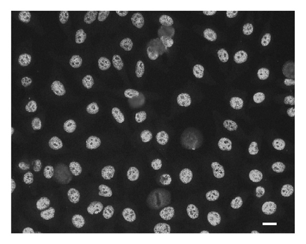
Figure 1.

Indirect immunofluorescence pattern of anti-C1/C2-positive patient serum on HEp-2 cells. Cells were incubated with diluted patient serum (1:160), and bound IgG was detected by fluorescence microscopy after incubation with fluoresceinylated rabbit anti-human IgG. Strongly positive, very regular, coarse nuclear staining that spares nucleoli is observed. Original magnification is 200×. Scale bar is 10 μm.
