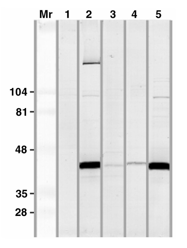
Figure 2.

Immunoblotting using different patient sera (2-5) and a normal control serum (1) diluted 1:100 on blots of HeLa-cell nuclear extracts separated on SDS-PAGE. The serum examined in lane 2 is identical to the one shown in Fig. 1, and the serum in lane 5 is a sample from the original patient described by Stanek et al [4]. Developed with anti-IgG antibodies. Molecular masses (×10-3) of standard proteins are indicated on the left.
