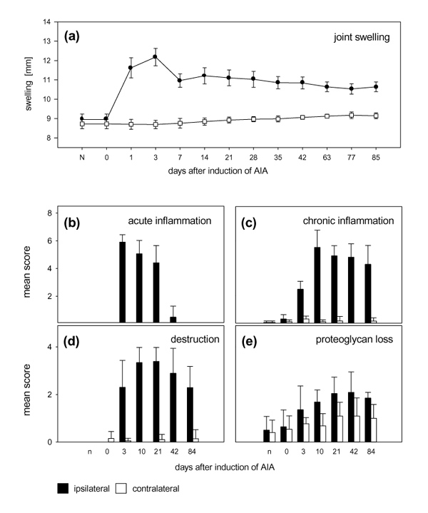
Figure 1.

Development of antigen-induced arthritis. (a) Diameters of the injected knee joints and the contralateral knee joints at different time points of arthritis. The contralateral knees show a slight increase in their diameters owing to growth. (bStuttgart: Gustav Fischer Verlag;d) Arthritic changes of the knee joints at 3, 10, 21, 42 and 84 days after the induction of arthritis, and of knee joints from immunized non-arthritic animals (0 days) in comparison with untreated normal control animals (n). (b) Histological examination of acute inflammatory changes (fibrin exudation and granulocytic infiltration of the synovial membrane and the joint cavity) in the injected joint and the contralateral knee. (c) Histological examination of the chronic inflammatory changes (hyperplasia of synovial lining, infiltration of mononuclear leucocytes) of the injected joint and the contralateral knee. (d) Overall joint destruction (pannus formation and erosion of cartilage and bone) in the injected knee joint and in the contralateral knee joint. (e) Loss of proteoglycans (staining with safranin O) in the cartilage of the injected knee and the contralateral knee. All values are means and standard deviations.
