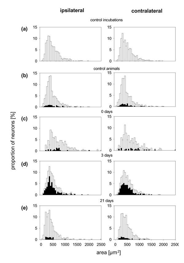
Figure 6.

Size distribution of the cultured neurons from different experimental groups. The white bars show the proportions of neurons with different areas from experiments in which SP–gold-binding sites were determined. The neurons with SP–gold-binding sites are shown by the black bars.
