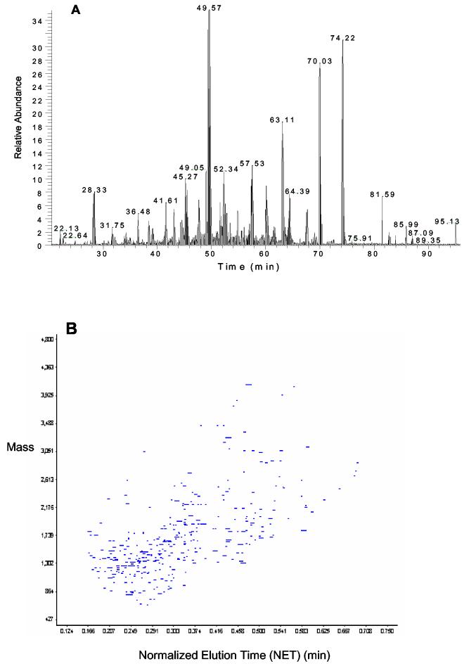
Figure 2.
(A) Base peak chromatogram of LTQ LC-MS/MS analysis of 5000 MCF-7 human breast cancer cells prepared using the TFE protocol. (B) 2-D plot of identified peptides from 5000 MCF-7 cancer cells using the TFE protocol. Peptides observed in the LC-FTICR analysis were displayed based on their monoisotopic masses and normalized elution times (NET).

