Figure 6.
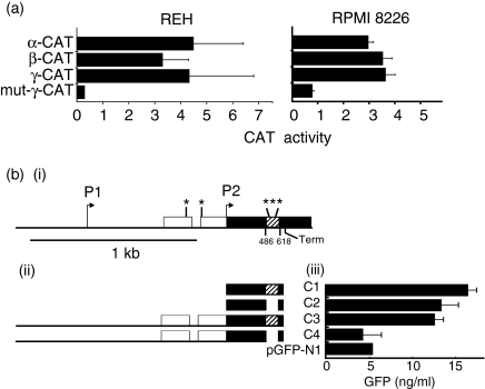
Enhancer activities of α, β and γ sequences. (a) Chloramphenicol acetyltransferase (CAT) assays. Left: activity of α, β and γ enhancer constructs in REH cells relative to that of the original pCAT-promoter vector. Right: activity of each construct co-transfected with pCMV-B-Myb into RPMI 8226 cells relative to the original pCAT-promoter vector co-transfected with pCMV-B-myb. (bi) The bcl-2 5′ noncoding sequences (white) and coding sequences containing the 132-bp sequence encompassing α, β and γ (black and cross-hatched respectively), cloned between the HCMV promoter and the green fluorescent protein (GFP) gene. Also indicated: Myb-binding sites (*), transcriptional start sites (arrows) and point of RNA chain termination (Term). (bii) Regions of the bcl-2 gene cloned into pGFP-N1 to produce constructs C1-4. (biii) Levels of GFP observed in REH cells transfected with constructs C1-4 or with the empty pGFP-N1 vector (pGFP-N1).

