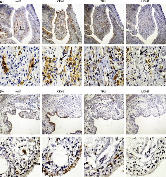
Figure 1.
Expression patterns of LIGHT and TR2 in RA synovium. Synovial tissue sections from RA (a) and OA patients (b) were analyzed for the expression of vWF (the marker for endothelial cells), LIGHT, TR2, and CD68 (the marker for macrophages) using immunohistochemical analysis as described in materials and methods. Sublining areas around the newly formed microvessels are magnified in the lower panel. The box in the low magnification picture (40X, upper panel) indicates the area magnified in high magnification pictures (400X, lower panel). Note that microvessels (arrows) are stained with antivWF antibodies but not with other antibodies. The pictures are representatives from analysis using 7 different RA and 5 different OA synovial tissue specimens.

