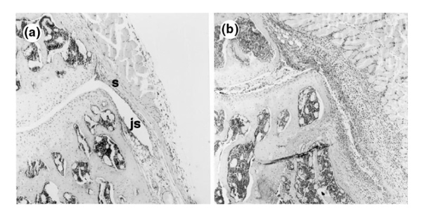
Figure 1.

Inflammation in H&E-stained sections of mouse knee joints after induction of immune-mediated arthritis (ICA). (a) Section from FcR γ-chain-/- mouse 3 days after ICA induction. No inflammatory cells are visible in the joint space (js) or synovium (s). (b) Section from C57BL/6 (control) mouse 3 days after ICA induction. Florid inflammation is visible both in the joint space (exudate) and in the synovium (infiltrate) (orginal magnification 100×).
