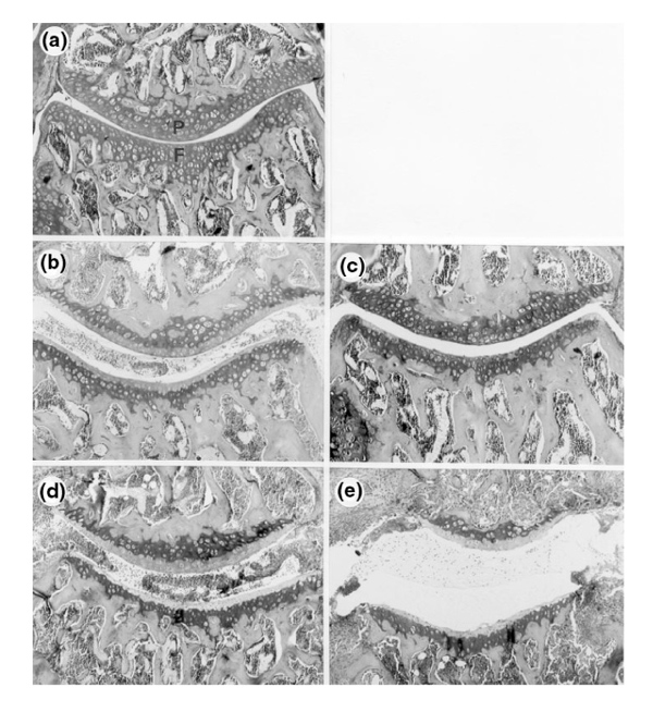
Figure 6.

Cartilage damage during ICA. Safranin-O-stained knee-joint sections of FcR γ-chain-/-, C57BL/6, and DBA/1 mice 3 and 7 days after induction of ICA. Proteoglycan depletion was correlated with destaining of the superficial layer of the cartilage matrix, which is normally stained red. Both patellar (P) and femoral (F) cartilage are shown. (a) FcR γ-chain-/- 3 days after ICA induction. No proteoglycan depletion is seen. (b) C57BL/6 mouse 3 days after ICA induction. Marked destaining of the matrix is found. (c) C57BL/6 mouse 7 days after ICA induction. The absence of depleted areas suggests that the matrix has been completely restored. (d) DBA/1 mouse 3 days after ICA induction. The cartilage matrix is completely depleted, indicating considerable PG loss. (e) Same mouse strain (DBA/1) as in (d), 7 days after ICA induction. The cartilage matrix seems still devoid of PG, suggesting that no repair has taken place. Moreover, marked erosion of the matrix is visible, mainly of the femoral cartilage.
