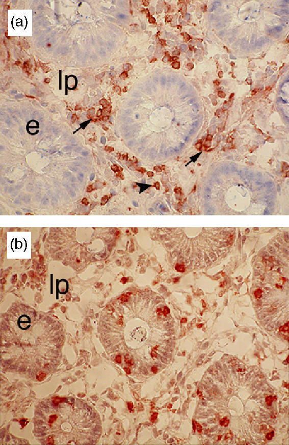
Figure 2.

Immunohistochemical staining of colonic sections obtained from a healthy pig with anti-CD4 (a) and anti-TCR-δ (b). Colonic CD4+ T cells were restricted to the lamina propria (lp) and distributed as either small clusters (black arrow) or isolated cells (arrowhead). γδ T cells were scattered throughout the lamina propria and epithelium (e). Original magnification: ×400.
