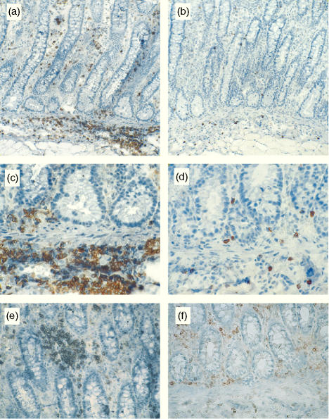
Figure 4.
Representative micrographs of CD4+ and γδ T-cell distribution at the colonic mucosa of pigs with swine dysentery (a–e). CD4+ T cells accumulated in large aggregates in the lamina propria (e) and across the submucosa (a,c). Small numbers of γδ T cells were also found in these LESions (b,d). CD4+ T cells were more evenly distributed in the colonic mucosa of control pigs (f).

