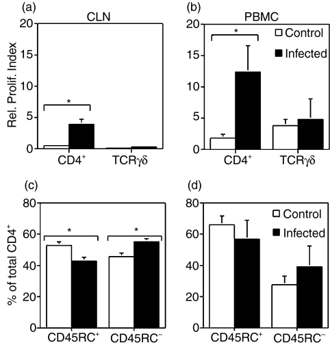
Figure 5.
Antigen-specific proliferation (a,b) and phenotype (c,d) of lymphocytes recovered from peripheral blood and colonic lymph nodes (CLN). PBMC and CLN were isolated on day 15 postchallenge and stained with PKH67 (a,b). Cells were cultured for 5 days with medium or Brachyspira hyodysenteriae antigens. After harvesting, cells were labelled with anti-CD4 or anti-TCR-γδ and analysed by flow cytometry. As cells divided, the PKH67 dye was split between daughter cells, and proliferation was measured by the decrease in PKH67 fluorescence intensity. A relative proliferation index was calculated by dividing the percentage of CD4+ or γδ T cells that proliferated in antigen-stimulated cultures by the percentage of CD4+ or γδ T cells that proliferated in cultures maintained with media alone (i.e. unstimulated). In addition, freshly isolated PBMC and CLN were double-stained with anti-CD4 and anti-CD45RC (c,d). The percentage of CLN CD4+ T cells with a memory/activated phenotype (i.e. CD4+ CD45RC−) was significantly increased in pigs that were challenged (c,d) (*P < 0·05). Flow cytometric analysis of CD45RC expression (c,d) was performed by gating only on CD4+ T cells.

