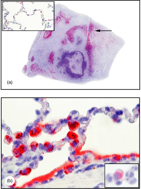
Figure 1.
Enhanced porcine surfactant protein D (pSP-D) immunoreactivity in chronic Actinobacillus pleuropneumoniae bronchopneumonia. (a) Strong pSP-D expression in lung tissue, with no pSP-D in fibrotic and necrotic areas (approximate magnification ×2). [The arrow indicates the area represented in (b). ] (b) Strong pSP-D immunoreactivity, diffuse in the surfactant lining alveoli and in hypertrophic alveolar type II cells, and distinguishable from alveolar macrophages by cytokeratin immunoreactivity (lower insert shown in panel b) (magnification ×250, oil). For comparison, in normal lung tissue pSP-D immunoreactivity is present in non-hypertrophic alveolar type II cells but not in the alveoli (upper insert) (magnification ×100). Detection was performed in the sections shown in panels (a) and (b) by using amino-ethyl-carbazole (AEC) and in the insert of (b) by using Fast red. All sections were counterstained with Mayers's haematoxylin.

