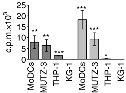
Figure 1.
Proliferation of CD4+ CD45RA+ T cells after stimulation of monocyte-derived dendritic cells (MoDCs) and differentiated MUTZ-3, THP-1 and KG-1 cell lines. The differentiated cell lines and MoDCs were cultured for 48 hr in the presence of lipopolysaccharide (LPS) (dark grey bars) or a proinflammatory cocktail (light grey bars) prior to co-culture with CD4+ CD45RA+ T cells for 4 days. The results shown represent net proliferation, i.e. proliferation induced by unstimulated cells is subtracted. The values shown are the average of four replicate cultures ± standard deviation (*P < 0·05; **P < 0·005; ***P < 0·0005). c.p.m., counts per minute.

