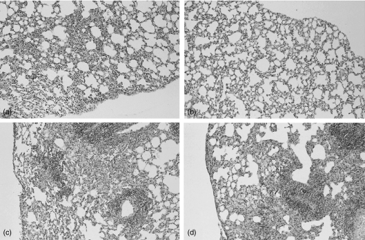
Figure 6.
Representative histologic sections of lungs of Wt (a and c) and p40 Tg mice (b and d) 2 (a and b) and 5 (c and d) weeks after inoculation with M. tuberculosis, demonstrating less inflammatory infiltrate in p40 Tg mice 2 week p.i. as compared to Wt mice. Haematoxylin and eosin staining, original magnification ×33.

