
Figure 1.
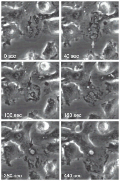
Time-lapse video-microscopy of macropinosome formation in murine bone-marrow-derived DC. The formation and fate of two macropinosomes formed at the leading edge of a DC cultured in the absence of growth factors for 24 hr prior to examination. At 0 seconds a macropinsome forms (black arrow) and subsequently (40–280 seconds) traffics towards the nucleus. At 40 seconds a large circular ruffle forms at the leading edge of the DC (white arrow), closes off to form a macropinosome (100 seconds), fuses with other large vesicles (180 seconds) and finally fuses with the initial macropinosome.