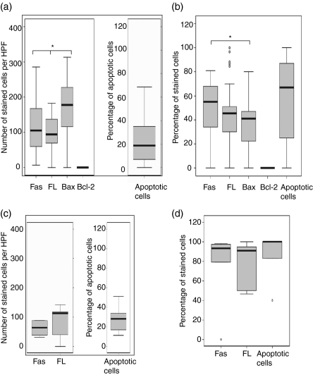
Figure 3.
In situ distribution of Fas ligand (FasL), Fas, Bcl-2, Bax and apoptosis in the tuberculosis (TB) (a,b) granulomas, and FasL, Fas, and apoptosis in foreign body (FB) (c,d) granulomas, analysed by using immunohistochemical staining. The median, 25th and 75th percentiles, and minimum and maximum values are shown. The marks indicate the extreme values. The Wilcoxon signed-rank test was used for matched analysis and the Mann–Whitney test for two independent group comparisons. Statistical significance with a P-value of <0·05 is marked with an asterisk. (a) Number of stained cells per high power field (HPF) in the epithelioid cells within TB granulomas. Bax-expressing cells were more numerous than either Fas (P = 0·01) or FasL-expressing cells. Bcl-2-expressing cells were very few or could not be detected in the granulomas. (b) Percentage of stained cells in multinucleated giant cells in TB granulomas. The percentage of giant cells expressing Bax was lower than those expressing Fas (P = 0·05). (c) Number of stained cells per HPF in the epithelioid cells of FB granulomas. There was no significant difference in the expression of cytokines. (d) Percentage of stained cells in multinucleated giant cells in FB granulomas. There was no significant difference in the expression of cytokines. Among TB giant cells there were significantly lower percentages of FasL and Fas and apoptotic cells as compared with FB giant cells.

