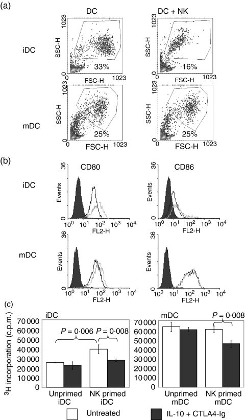
Figure 4.
Natural killer (NK) cells modified allogeneic dendritic cell (DC) function. Immature and mature DCs (iDCs and mDCs, respectively) were co-cultured with allogeneic NK cells at a ratio of 5 : 1 for 3 days. (a) Gated histograms represent the forward side-scatter profile of DCs and reveal a significant decrease in iDC numbers after culture with NK cells. (b) Flow cytometric analysis was performed on gated DCs using monoclonal antibodies (mAbs) directed against CD80 and CD86. The expression of CD80 and CD86 on DCs cultured alone (black line) or DCs co-cultured with NK cells (grey line) is shown in the histogram and demonstrates up-regulation of CD80 on iDCs. The shaded histogram represents isotype-matched control mAb staining. NK cells were distinguished from DCs and excluded from analysis by gating based on their forward/side-scatter profiles. Data shown are representative of three independent experiments. (c) After 3 days of culture with allogeneic DCs, NK cells were removed by aspiration and density gradient separation. Viable DCs were counted and used as stimulators of allogeneic CD4+ T cells in the absence of NK cells. MLR stimulated by DCs, which were precultured with NK cells, was sensitive to the inhibitory effects of suboptimal CTLA4-Ig/interleukin (IL)-10. Proliferation was measured by [3H] thymidine incorporation and results are expressed as the percentage inhibition of proliferation in comparison with untreated controls. P-values were determined by unpaired Student's t-test.

