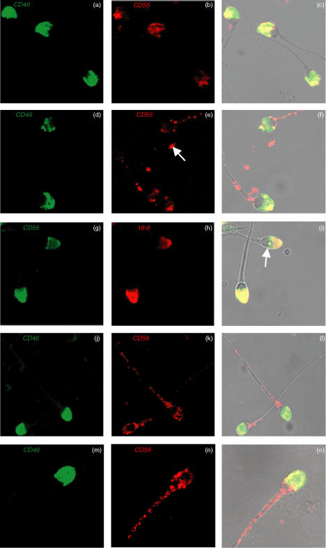
Figure 2.
CLSM images of dual immunofluorescent localisation of CD46 (green) & CD55 (red) on prefix (a–c) & postfix (d–f) AR-spermatozoa. Arrow indicates small structures not associated with intact spermatozoa (e). CD55 (green) & 18-6 mAb (red) on prefix AR-spermatozoa (g–i); arrow indicates spermatozoal equatorial segment (i). CD46 (green) & CD59 (red) expression on both prefix (j–l) and postfix (m–o) AR-spermatozoa. Co-localisation signals as yellow pixels for CD46 & CD55 (c & f), CD55 & 18–6 mAb (i) and CD46 & CD59 (l&o). Final magnification: × 1200 (a–i), × 1000 (j–l) and ×1500 (m–o).

