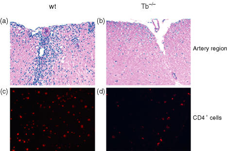
Figure 4.
Infiltration of leucocytes into the CNS of T-bet−/− (Tb−/−) and wild type (wt) littermates. Animals were killed on day 15 at the peak of disease, and spinal cords were fixed (10% buffered formalin) and infiltration was assessed by H&E staining and visualized at ×10 for wild type (a) and T-bet−/− (b) mice in the arterial region. The tissue sections were stained for the CD4+ T cells in wild-type (c) and T-bet−/− mice (d) Data shown are representative of eight different animals.

