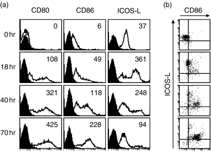
Figure 2.
PDC mature in the presence of antigen-experienced T cells. (a) Freshly isolated PDC were cocultured with peripheral blood CD4+ CD45RO+ T cells for the indicated time periods in the presence of SEB and analysed by flow cytometry for the expression of CD80, CD86 and ICOS-L. Black histograms represent isotype control staining, the inset numbers indicate the mean-fluorescence over all cells minus the mean fluorescence of the isotype control. Shown is one representative experiment out of three. (b) Two-colour staining of the PDC for the expression of CD86 and ICOS-L. One representative experiment out of three is shown.

