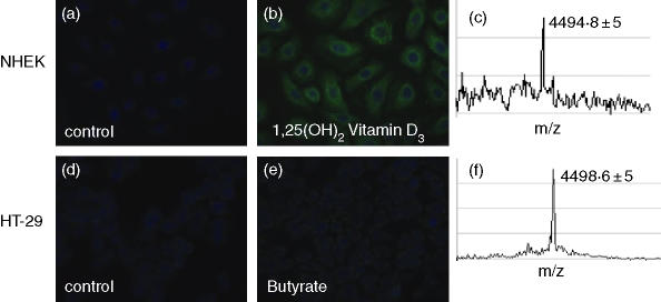
Figure 2.
Cathelicidin peptide expression in keratinocytes and colon epithelial cells. (a–c) NHEK grown on chamber-slides were stimulated with the vehicle, or 1,25(OH)2 VD3; (d–f) colonocytes (HT-29) were stimulated with the vehicle or butyrate. Cells were stained with a polyclonal anti-LL-37 antibody and nuclei were detected with DAPI in (a,b,d,e). Immunofluorescence is displayed at 400× magnification. Processing to active cathelicidin peptide was evaluated by SELDI-TOF analyses of NHEK and HT-29 cells in (c) and (f) after stimulation with VD3 or butyrate, respectively. A peak at 4 494 Da corresponding to the mature LL-37 peptide was detected in NHEK and HT-29 cells. Data from one representative of three independent experiments are displayed.

