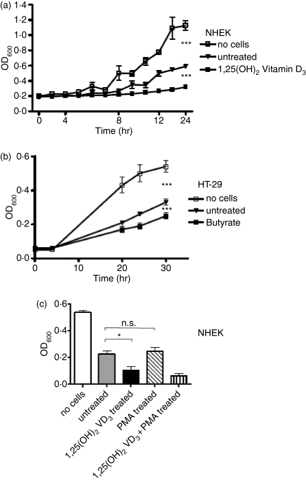
Figure 7.
Antimicrobial activity of stimulated keratinocytes and colon epithelial cells. (a) NHEK were stimulated with 1,25(OH)2 VD3 for 24 hr, cells were harvested and cell lysates were coincubated with S. aureusΔmprF and bacterial growth was monitored over time to determine antimicrobial activity. Conditions not containing cell lysates or containing cell lysates from unstimulated cells were used as controls. (b) HT-29 colon cells were stimulated with butyrate for 24 hr and cell lysates were incubated with S. aureusΔmprF. (c) NHEK were stimulated with 1,25(OH)2 VD3, or PMA to induce HBD2 expression and cell lysates were incubated with S. aureusΔmprF. Bacterial growth was measured by OD600. All data shown are means (± SD) of triplicates. (*P < 0·05; **P < 0·01; Student's t-test).

