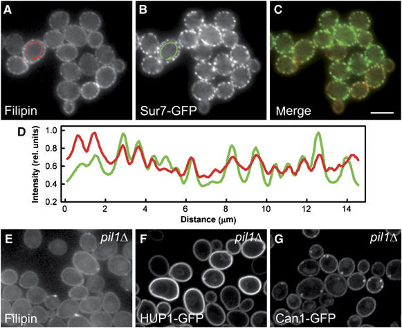
Figure 1.

Sites of sterol accumulation in plasma membrane colocalize with MCC. Simultaneous localization of filipin-stained sterols (A; red in C, D) and an MCC marker Sur7GFP (B; green in C, D) was performed in living GYS48 cells. Wide-field fluorescence micrographs (A–C) and the fluorescence intensity profiles along the cell surface (D; outside the arrows in A, B) are presented. The curves in (D) were smoothed using a mean filter to reduce the noise and normalized to the same maximum value. Distribution of filipin-stained sterols in pil1Δ mutant (GYS130; E) was compared with HUP1 (F) and Can1p (G) patterns in these cells (strains GYS131 and GYS132, respectively). Again, wide-field image is presented in (E), whereas confocal sections are shown in (F, G). Bar: 5 μm.
