Figure 2.

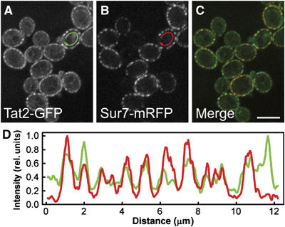
Tat2p is a part of MCC. Simultaneous localization of the tryptophan permease Tat2GFP (A; green in C) and the MCC marker Sur7mRFP (B; red in C) was performed in living GYS122 cells. Fluorescence intensity profiles along the cell surface (D; outside the arrows in A, B; green: Tat2GFP; red: Sur7mRFP) are presented. The curves in (D) were smoothed using a mean filter to reduce the noise and normalized to the same maximum value. Bar: 5 μm.
