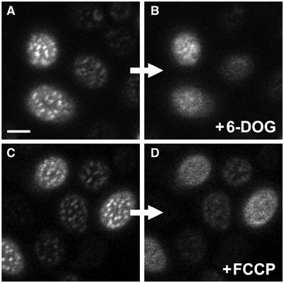
Figure 3.

HUP1 patches are dissolved after plasma membrane depolarization. Changes in the distribution of HUP1GFP in the plasma membrane of living cells depolarized by 6-DOG uptake (strain GYS118; A, B) or FCCP treatment (strain GYS110; C, D; see Materials and methods for details) were observed. Surface confocal sections of the same cells before (A, C) and after the treatment (B, D) are presented. In both cases, the protein was released from MCC patches after plasma membrane depolarization. Only remnants of patches are visible in some treated cells. Bar: 2 μm. For corresponding time-lapse observations, see Supplementary Movies S1, S2 and S3.
