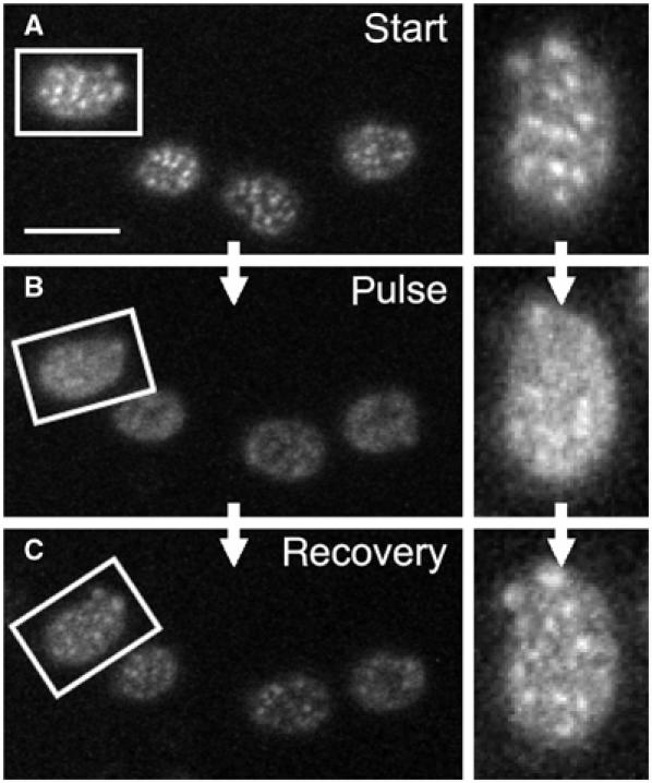
Figure 4.

Depolarization-induced changes in HUP1 distribution are reversible. For membrane depolarization, 1 min pulse of external electric field was applied on exponentially growing cells expressing HUP1GFP (strain GYS110) in order to depolarize the plasma membrane. The distribution of the protein before (A), immediately after (B) and 20 min after depolarization (C) is shown. Aligned surface sections of one cell are also presented (right). Despite the increased signal-to-noise ratio caused by phototobleaching of HUP1GFP fluorescence during the scanning, the restored pattern of MCC patches almost identical to that in (A) is clearly visible in (C). Bar: 5 μm.
